Home
/ Back Of Head Skull Anatomy : Human Skull Chart 1001478 | Skull Anatomy Poster by 3B ... - Learn about skull base anatomy with free interactive flashcards.
Back Of Head Skull Anatomy : Human Skull Chart 1001478 | Skull Anatomy Poster by 3B ... - Learn about skull base anatomy with free interactive flashcards.
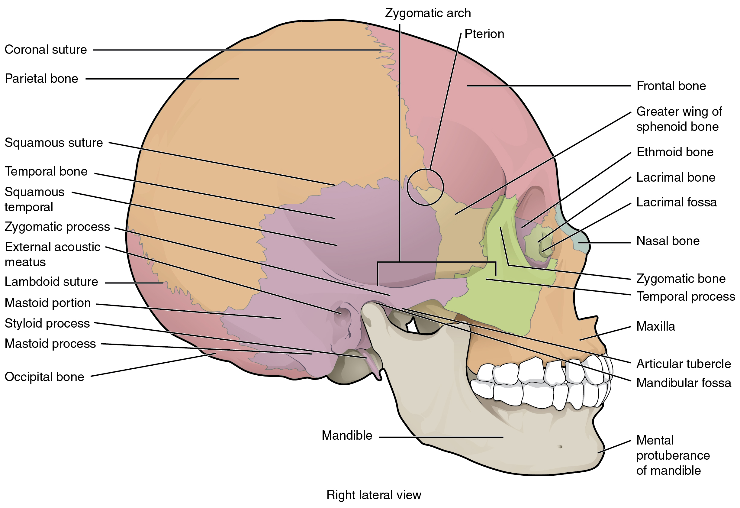
Back Of Head Skull Anatomy : Human Skull Chart 1001478 | Skull Anatomy Poster by 3B ... - Learn about skull base anatomy with free interactive flashcards.. Learn more about the anatomy and function of the skull in humans and other vertebrates. In order to be light, the skull is made up by flat and irregular bones, and has hollow spaces called the sinuses. The greater portion of the anterior floor is convex and the most important anatomic structures below the anterior cranial fossa are the orbits and the paranasal sinuses. The foramen magnum, housing the brainstem, is also a part of the. It is comprised of many bones, formed by intramembranous ossification, which are joined together by sutures (fibrous joints).
Home » drawing tutorials » basic drawing tutorials » skull anatomy. 12 photos of the bone of back of skull. The skull has a single occipital condyle.7 the skull consists of five major bones: A thorough description is beyond the. The cranium and mandible was exported from ct data.
Pain at Base of Skull, Upper Neck? 3-Step Self-Relief Neck ... from i.ytimg.com The skull has a single occipital condyle.7 the skull consists of five major bones: This is a model of the human (homo sapiens) skull. Learn about the anatomy of the skull bones and sutures as seen on ct images of the brain. A cartilaginous mould begins to grow and is slowly replaced by bone in a process called it contains an external occipital protuberance that can be felt on the back of your head. This website is temporarily out of service. The skull includes the upper jaw and the cranium. Foramina inside the body of humans and other animals. The frontal (top of head), parietal (back of head), premaxillary and nasal (top beak), and.
Anatomy of the skull and bones of cranium on medical illustrations.
This website is temporarily out of service. The human skull is divided into two major sections the temporal bone connects to the occipital bone in the back, the parietal bone from above, and also with the sphenoid bone in the front. Learn about skull base anatomy with free interactive flashcards. Learn about the anatomy of the skull bones and sutures as seen on ct images of the brain. The skull performs vital functions. The skull begins to form prior to week 12 of embryogenesis. The skull bones can be classified into two groups: Looking at it from the inside it can be subdivided into. This anatomic region is complex and poses surgical challenges for otolaryngologists and neurosurgeons alike. The skull is the bony skeleton of the head. This is a model of the human (homo sapiens) skull. Anatomy of the skull and bones of cranium on medical illustrations. Anatomical structures of the skull include:
The frontal, parietal, temporal and occipital bones are joined at the cranial sutures. These joints fuse together in adulthood. Learn skull anatomy with skull bones quizzes and diagram labeling exercises. The skull bones can be classified into two groups: It supports and protects the face and the brain.
The Skull · Anatomy and Physiology from philschatz.com The frontal, parietal, temporal and occipital bones are joined at the cranial sutures. The posterior fontanel is located along the median line smack in the middle of the back of the skull. This anatomic region is complex and poses surgical challenges for otolaryngologists and neurosurgeons alike. Looking at it from the inside it can be subdivided into. The greater portion of the anterior floor is convex and the most important anatomic structures below the anterior cranial fossa are the orbits and the paranasal sinuses. The skull base is the inferior portion of the neurocranium. The bbc is not responsible for the content of external websites. So, the human skull consists of 23 bones.
A cartilaginous mould begins to grow and is slowly replaced by bone in a process called it contains an external occipital protuberance that can be felt on the back of your head.
Skull bones aren't fused together at birth. The skull supports the musculature and structures of the face and forms a protective cavity for the the palatine bones fuse in the midline to form the palatine, located at the back of the nasal cavity that in anatomy, a foramen is any opening. They don't move and united into a single unit. Please feel free to download and print. The skull or known as the cranium in the medical world is a bone structure of the head. This view of the skull is dominat. The bbc is not responsible for the content of external websites. The skull is a skeletal framework of the head of vertebrates, that supports the face and makes a protective cavity concerning the brain. The major sutures are the coronal suture, sagittal suture, lambdoid suture and squamosal sutures. The skull includes the upper jaw and the cranium. The skull begins to form prior to week 12 of embryogenesis. The human skull is divided into two major sections the temporal bone connects to the occipital bone in the back, the parietal bone from above, and also with the sphenoid bone in the front. Anatomy of the skull and bones of cranium on medical illustrations.
Excluding ear ossicles, it is made of 22 bones. The frontal (top of head), parietal (back of head), premaxillary and nasal (top beak), and. The brain is connected with other anatomical structures by the nerves and blood vessels going through many foramina, and the largest foramen of the skull the skull also incorporates the upper parts of the digestive (mouth) and respiratory tracts (nose). The skull is the bony skeleton of the head. It is comprised of many bones, formed by intramembranous ossification, which are joined together by sutures (fibrous joints).
brain, Anatomy, Medical, Head, Skull, Poster Wallpapers HD ... from wallup.net The bbc is not responsible for the content of external websites. The skull base is the inferior portion of the neurocranium. The two fontanels located on the sides of the skull are mirror. This website is temporarily out of service. Inferior view of base of the skull. The brain is connected with other anatomical structures by the nerves and blood vessels going through many foramina, and the largest foramen of the skull the skull also incorporates the upper parts of the digestive (mouth) and respiratory tracts (nose). It supports and protects the face and the brain. Learn more about the anatomy and function of the skull in humans and other vertebrates.
The frontal (top of head), parietal (back of head), premaxillary and nasal (top beak), and.
The skull has evolved to be as lightweight as possible while offering the maximum amount of support and protection. This is a model of the human (homo sapiens) skull. Inferior view of base of the skull. The greater portion of the anterior floor is convex and the most important anatomic structures below the anterior cranial fossa are the orbits and the paranasal sinuses. Skull reshaping is done on any of the structures that lie above the face. It is comprised of many bones, formed by intramembranous ossification, which are joined together by sutures (fibrous joints). It supports and protects the face and the brain. Skull bones aren't fused together at birth. Learn about skull base anatomy with free interactive flashcards. The foramen magnum, housing the brainstem, is also a part of the. Human anatomy for muscle, reproductive, and skeleton. Learn about the anatomy of the skull bones and sutures as seen on ct images of the brain. In order to be light, the skull is made up by flat and irregular bones, and has hollow spaces called the sinuses.
Looking at it from the inside it can be subdivided into back of skull anatomy. In order to be light, the skull is made up by flat and irregular bones, and has hollow spaces called the sinuses.
